[最も好ましい] 1 wall bony defect 772812-1 wall bony defect
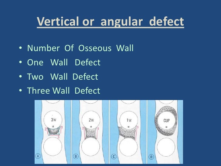
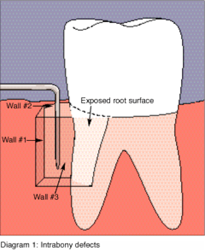
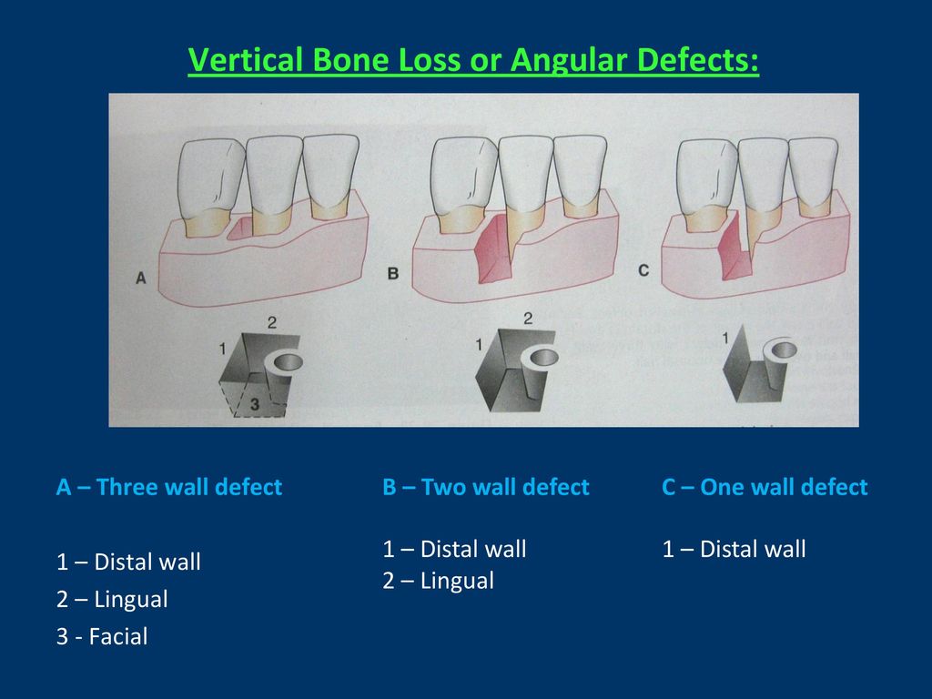
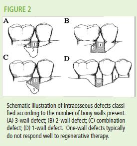
A tiny bony defect is seen in the posterior wall of the right frontal sinus, on the right side of the crista galli, measuring 15 mm in diameter This tiny bony defect allows the contrast material to enter the right frontal sinus as seen on image #65, series #602 Less dense contrast material is seen filling the left frontal sinus1 J Periodontol 07 May;78(5)8 Favorable periodontal healing of 1wall infrabony defects after application of calcium phosphate cement wall alone or in combination with enamel matrix derivative a pilot study with canine mandibles Shirakata Y(1), Yoshimoto T, Goto H, Yonamine Y, Kadomatsu H, Miyamoto M, Nakamura T, Hayashi C, Izumi YAngular defects are classified on the basis of the number of osseous walls1 \ngular defects mas have one, two, or three walls digs 2315 to 2317) The number of walls in the apical portion of the defect may be greater than that in its occlusal portion, in which case the term combined osseous defect is used d ig 2318)

Periodontal Disease Wikipedia
1 wall bony defect
1 wall bony defect-The bone defects formed in this way are defined as a deformity or concavity comprising the alveolar bone and may involve one or more teeth Bony defect occurring in oblique direction alongside the root surface and having a base located in the apical of the alveolar crest is called 'infrabony' or 'intraosseous' defectThe cortical bone graft fitted in the outer attic wall bony defect In 18 patients, glass ionomer bone cement (GIBC) was used for further fixation of the cortical bone graft The GIBC (Medicem;


Classification Scheme For Intrabony Defects Figure 1 Reproduced Download Scientific Diagram
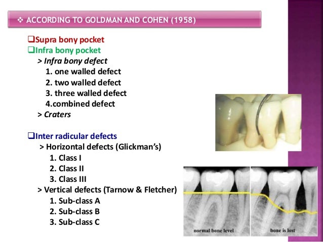
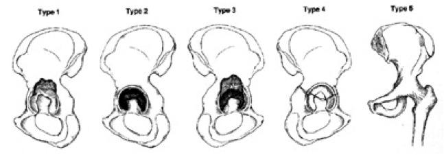
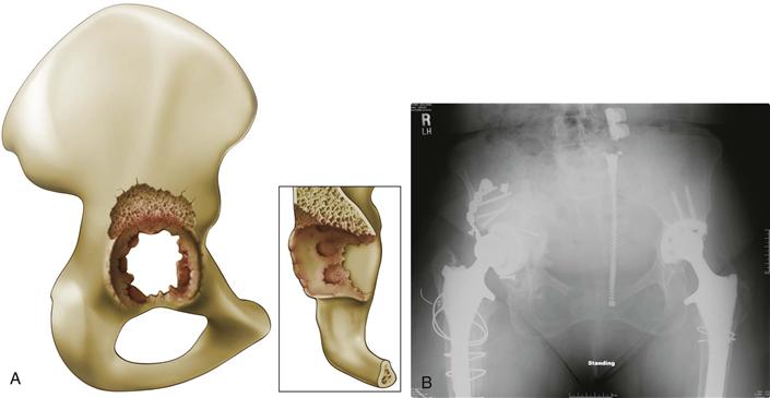
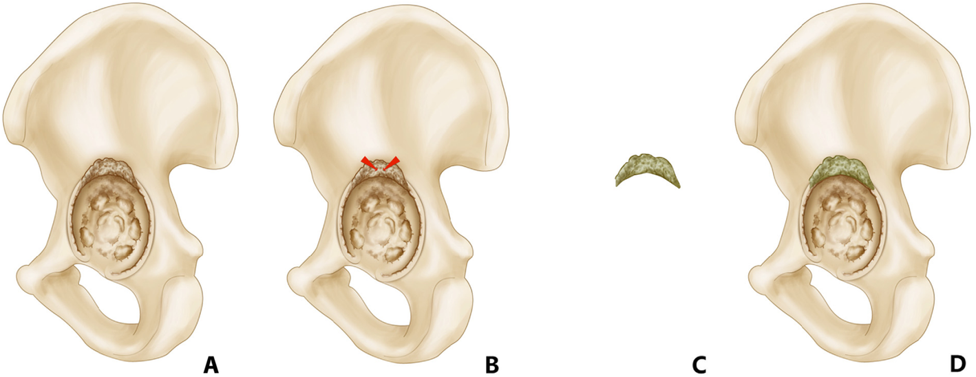
This patient had angle's class 3 malocclusion with anterior crossbite, further presenting some crowding with lack of right maxillary space This patient refeA central segmental defect involves loss of the medial wall of the acetabulum A cavitary deficiency (type II) is defined as volumetric bony loss of the acetabulum with an intact rim These defects can occur peripherally or centrally Medial cavitary defects involve loss of bone centrally with an intact medial wall, as in most cases of protrusioAn intrabony defect therefore can be sub classified, with respect to the number of remaining bony walls, in three categories the 1wall, 2wall and 3wall defects (Goldman and Cohen, 1958) The furcation involvements may also be included in the group of periodontal bony defects
7 (175%) at the middle third;The adjunctive use of DBBM in predominantly onewall defects seemed to compensate for, at least in part, the unfavorable osseous characteristics in terms of the outcomes of the procedure ViewThe size of the 40 bone defects ranged from 19 to 135 mm in width (mean, 66 mm) and from 11 to 50 mm in depth (mean, 23 mm) The bone defects were most commonly located at the lateral third of the posterior wall of the temporal bone 33 (5%) were located at the lateral third;
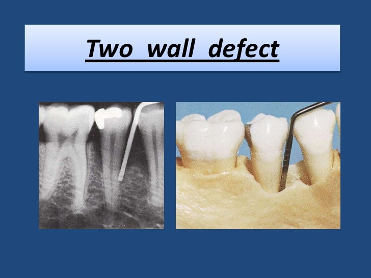
Promedica Dental Material, Neumunster, Germany) was prepared by mixing the powder with the provided solvent in the recommended ratio (11) 15 , 18Of onewall intraosseous bony defect of more than 4mm depth and 37o defect angle Though there are indications that two and three walled bony defects respond better to regenerative therapy than onewall defects, several studies have demonstrated that the extent of vertical attachment gain 1314 or osseous filling 13, 1516 corre1 the most common bony defect 2 located interdentally 3 consist of facial bony wall lingual bony wall two adjacent roots Notes on Twowall infrabony defects Also called an Intrabony defect Want to get rid of most, if not all, of the inflammation before performing surgery!


Bone Loss


Evaluation Of The Prevalence And Distribution Of Bone Defects Associated With Chronic Periodontitis Using Cone Beam Computed Tomography A Radiographic Study
Anomalies of the lower limbs clubfoot;Periodontal regeneration in a onewall intrabony defect is a challenging and complex phenomenon The combination therapy of commercially available bone grafts with the innovative tissue engineering strategy, the platelet rich plasma, has emerged as a promising grafting modality for two and three walled intrabony osseous defects1 the most common bony defect 2 located interdentally 3 consist of facial bony wall lingual bony wall two adjacent roots Notes on Twowall infrabony defects Also called an Intrabony defect Want to get rid of most, if not all, of the inflammation before performing surgery!


Www Codsjod Com Doi Cods Pdf 10 5005 Jp Journals 0028


Ijms Free Full Text The Fate Of Transplanted Periodontal Ligament Stem Cells In Surgically Created Periodontal Defects In Rats Html
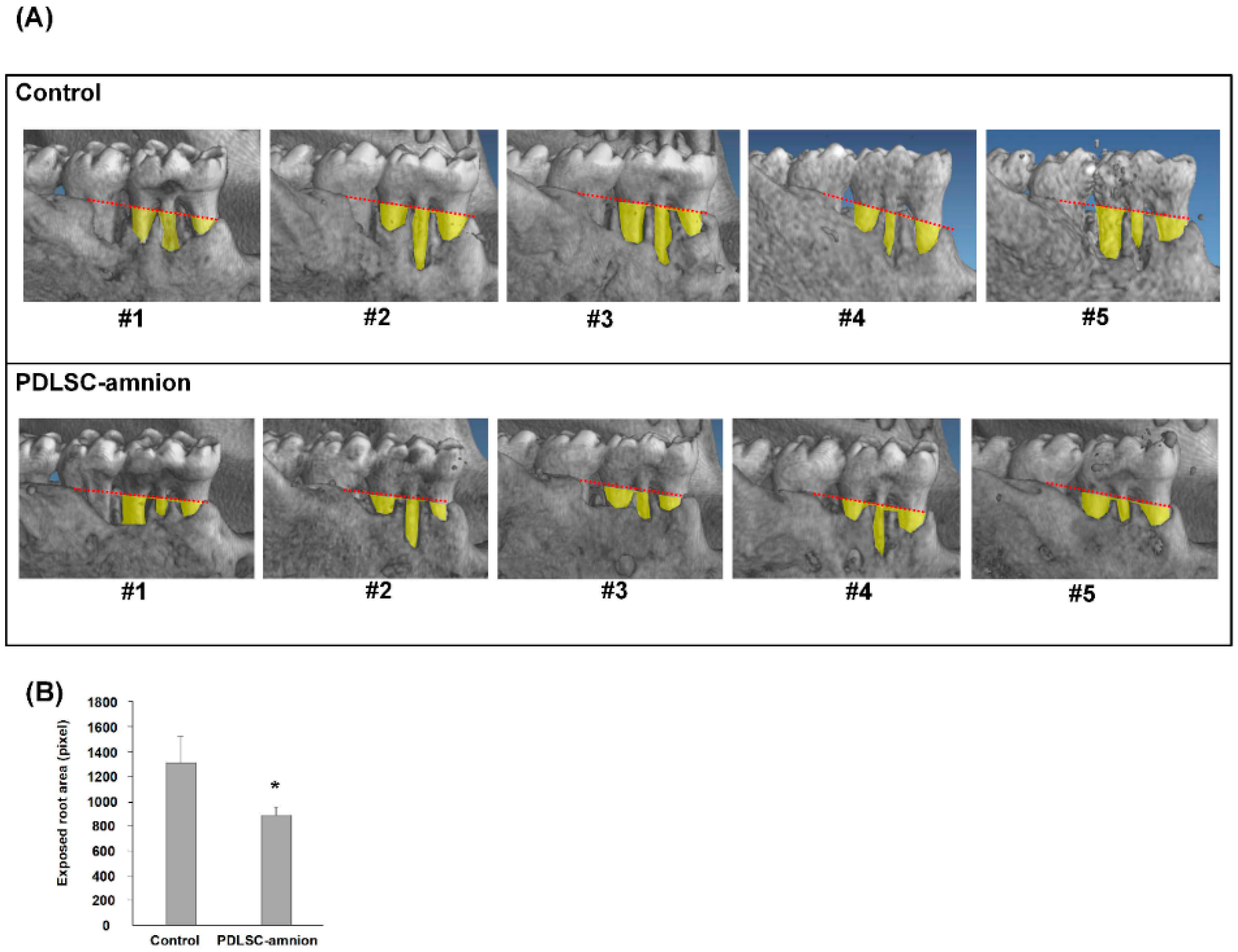
And none at the medial thirdMethods Onewall intrabony periodontal defects were surgically created at the distal aspect of the second and the mesial aspect of the fourth mandibular premolars in either right or left jaw1 J Periodontol 05 Aug;76(8) Treatment of wide, shallow, and predominantly 1wall intrabony defects with a bioabsorbable membrane a randomized controlled clinical trial Aimetti M(1), Romano F, Pigella E, Pranzini F, Debernardi C


Improved Long Term Treatment Outcomes Of Teeth With Deep Pockets And Reduced Periodontal Support By Periodontal Regenerative Surgery Oral Health Group


Patterns Of Bone Destruction In Periodontics
Papapanou PN, Wennström JL The angular bony defect as indicator of further alveolar bone loss J Clin Periodontol 1991;18(5)317–322 44 Greenstein B, Frantz B, Desai R, Proskin H, Campbell J, Caton J Stability of treated angular and horizontal bony defects a retrospective radiographic evaluation in a private periodontal practiceInfrabony defects With 1, 2 or 3wall bony defects Infrabony defect surrounded by 1, 2 or 3bony walls remaining The statistical analysis was performed using Chisquare test P < 005 was considered as statistically significant difference = O E E 2 χ2 ∑ Where, O=Observed number, E=Expected number RESULTSThoracic wall defect / thoracoschisis ectopia cordis;


Ossbuilder Titanium Membrane Implantology Osstem Uk


Bone Defect Repair On The Alveolar Wall Of The Maxillary Sinus Using Collagen Membranes And Temporal Fascia An Experimental Study In Monkeys
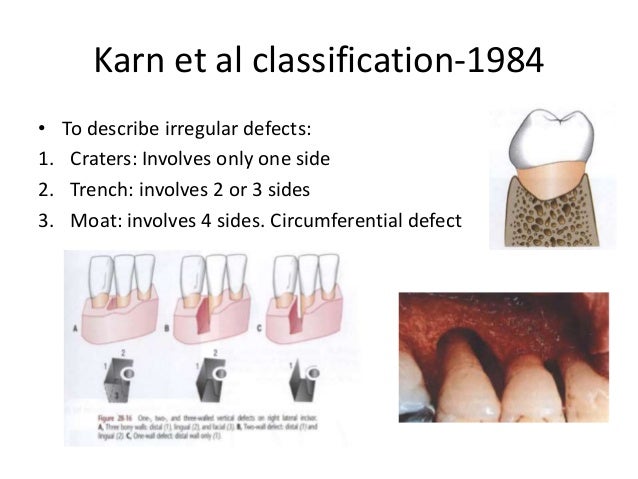
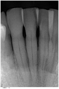
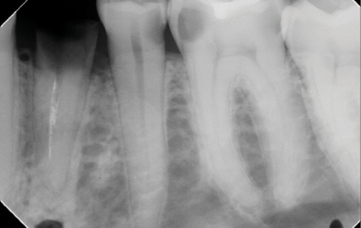
The bone destruction patterns that occur as a result of periodontal disease generally take on characteristic forms This Xray film displays a horizontal defect This Xray film displays two lonestanding mandibular teeth, #21 and #22 the lower left first premolar and canine, exhibiting severe bone loss of 3050%Osseous or Bony defects Any concavity or deformity in the alveolar bone that represents a change from normal contour as a result of periodontal disease EX craters, vertical defects (1 wall, 2 wall, 3 wall), hemiseptal, circumferential (moat)As 3D printing technology emerge, there is increasing demand for a more customizable implant in the repair of chestwall bony defects This article aims to present a custom design and fabrication method for repairing bony defects of the chest wall following tumour resection, which utilizes threedimensional (3D) printing and rapidprototyping technology


Evaluation Of Simulated Periodontal Defects Via Various Radiographic Methods Semantic Scholar


Jpis Journal Of Periodontal Implant Science
An intrabony defect therefore can be sub classified, with respect to the number of remaining bony walls, in three categories the 1wall, 2wall and 3wall defects (Goldman and Cohen, 1958) The furcation involvements may also be included in the group of periodontal bony defectsMethods Forty (39 evaluable) 3wall intrabony defects, each with a depth > or = 4 mm measured from the crest of the bony defect, were treated in 40 subjects with advanced chronic periodontitis Regeneration of angular bone defects was induced using nonresorbable membranes (GTR group;One or more sites with 1) a probing depth of 6mm or greater (>6mm), 2) a radiographic bony defect depth of greater than 3mm (>3mm), 3) sufficient keratinized tissue to allow complete coverage of the defect with gingival flaps, and 4) radiographic base of bony defect at least 2mm coronal to the apex of the tooth


1


Http Www Dental Campus Com Uploads Geistlichpdf En Is Cortellini Periodontal Regeneration Pdf
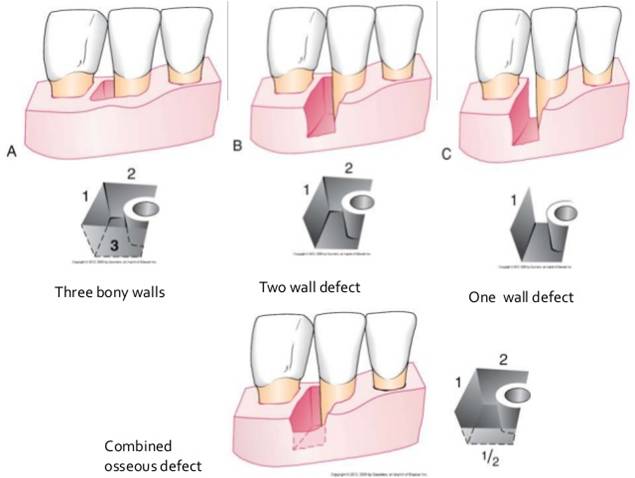
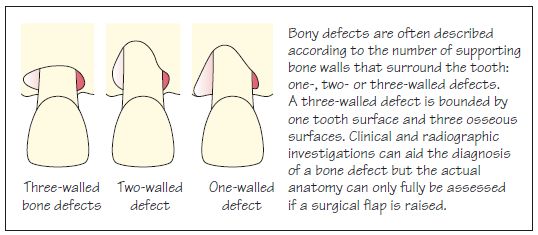
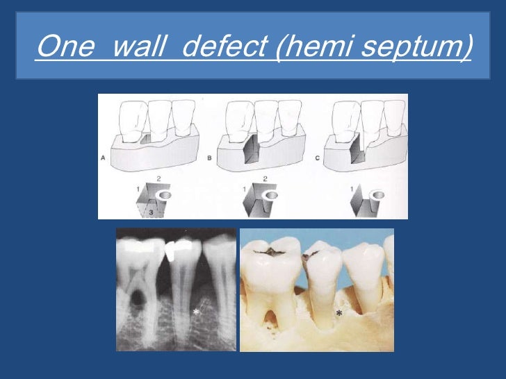
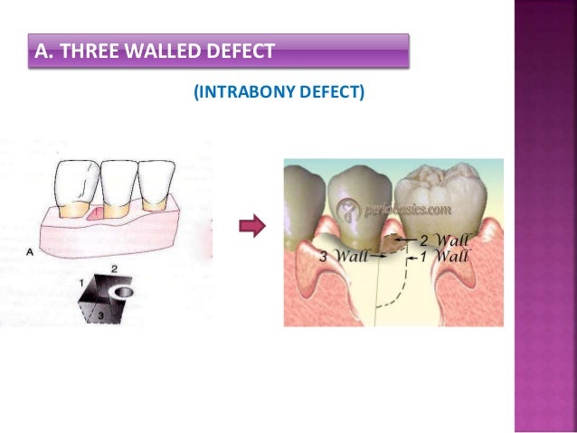
These bone defects are called bone defects A Infrabony Defects 1 Infrabony defects when bone resorption occurs unevenly, an oblique direction In infrabony defects of bone resorption primarily affects one tooth 2 Classification Infrabony defects Infrabony defects are classified based on the number of bone wallBone defects around the implant can manifest in various forms and affect the stability of the implant 13,17 Bone defects in periimplantitis lesions can be classified according to the criteria presented by Goldman and Cohen 18 Onewall intrabony defects are defects limited by 1 osseous wall and the implant surface Threewall intrabonyIf the defect is lined by only one wall of bone, then it is known as a onewall defect Bone defects which have the best prognosis for bone fill are threewall defects The increased number of walls and the height of these walls lends an increased bone matrix on which new bone can grow


Bone Loss Patterns


Treatment Of A Large Osseous Defect At Time Of Extraction And Implant Placement Inside Dentistry
Evaluable n = 19)The size of the 40 bone defects ranged from 19 to 135 mm in width (mean, 66 mm) and from 11 to 50 mm in depth (mean, 23 mm) The bone defects were most commonly located at the lateral third of the posterior wall of the temporal bone 33 (5%) were located at the lateral third;An intrabony defect therefore can be sub classified, with respect to the number of remaining bony walls, in three categories the 1wall, 2wall and 3wall defects (Goldman and Cohen, 1958) The furcation involvements may also be included in the group of periodontal bony defects


Www Ecronicon Com Ecde Pdf Ecde 18 Pdf
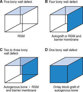


37 Tooth Extraction Socket Grafting And Barrier Membrane Bone Regeneration Pocket Dentistry
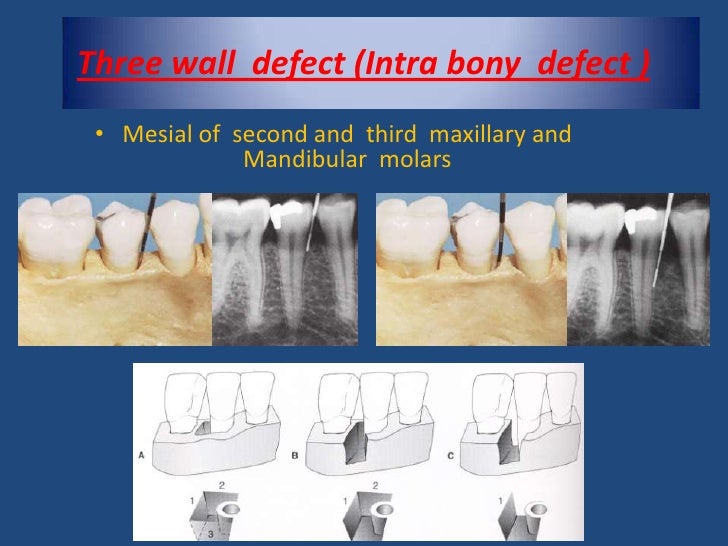
7 (175%) at the middle third;Diseases such as bone fracture and osteoporosis24 With reconstructive surgery, the buccal and lingual Huh et al21 reported that a safflower seed fraction mucoperiosteal flaps were elevated, and 1wall intra extract stimulates the formation of calcification nod bony defects (4 × 4 mm) were surgically created with ules, and has an increasingBone defects which have the best prognosis for bone fill are threewall defects The increased number of walls and the height of these walls lends an increased bone matrix on which new bone can grow When there are fewer numbers of bony walls associated with a shallow defect, the prognosis for success is decreased because the amount of


Figure 1 From A Canine Model For Histometric Evaluation Of Periodontal Regeneration Semantic Scholar


Lysostaphin And Bmp 2 Co Delivery Reduces S Aureus Infection And Regenerates Critical Sized Segmental Bone Defects Science Advances
Tal wound healing in intrabony defects in monkeys Materials and Methods Chronic twowall intrabony defects were created at the distal aspect of eight teeth in three monkeys (Macaca fascicularis) The 24 defects were randomly assigned to one of the following treatments (i) open flap debridement•The base of the defect is located apical to the surrounding bone •In most instances, angular defects have an accompanying intrabony periodontal pockets 10 Classified based on the number of osseous walls (Goldman & Cohen) 1 ONE WALLED DEFECTS 2 TWO WALLED DEFECTS 3 THREE WALLED DEFECTS 4 COMBINED OSSEOUS DEFECTS 11 A1 J Periodontol 05 Aug;76(8) Treatment of wide, shallow, and predominantly 1wall intrabony defects with a bioabsorbable membrane a randomized controlled clinical trial Aimetti M(1), Romano F, Pigella E, Pranzini F, Debernardi C


Periodontitis Pocket Dentistry
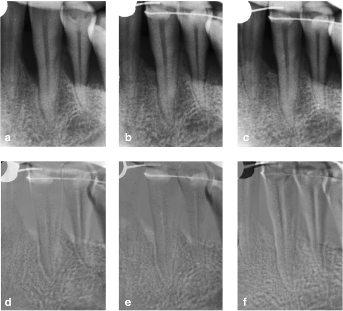


Treatment Of Intrabony Defects With Modified Perforated Membranes In Aggressive Periodontitis Subtraction Radiography Outcomes Prognostic Variables And Patient Morbidity Springerlink
Methods Onewall intrabony periodontal defects were surgically created at the distal aspect of the second and the mesial aspect of the fourth mandibular premolars in either right or left jawN = ) or EMD (EMD group;Dentaltown Where The Dental Community Lives®


Pathophysiology Of Periodontal Disease Revise Dental


Q Tbn And9gcqzjpi1sjuxtozyjgeneeswne G4nbynovkme8o65fijaja 5t Usqp Cau
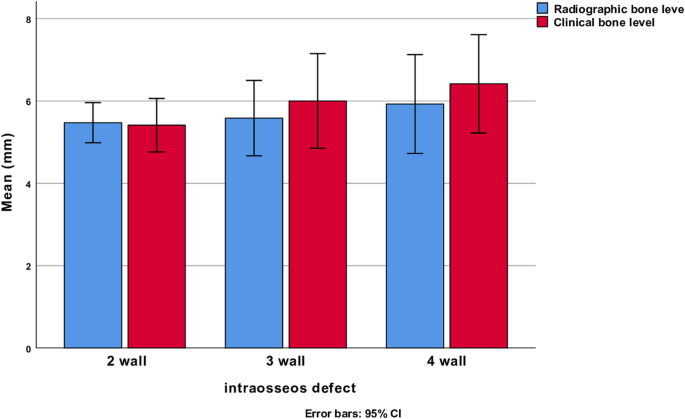
The distribution of location and defect types for implants with 2, 3, or 4wall bone defects are presented (Table 1) Data on clinical assessments performed immediately before and during surgical procedures are presented for defects located in the maxilla or the mandible (Table 2)A central segmental defect involves loss of the medial wall of the acetabulum A cavitary deficiency (type II) is defined as volumetric bony loss of the acetabulum with an intact rim These defects can occur peripherally or centrally Medial cavitary defects involve loss of bone centrally with an intact medial wall, as in most cases of protrusioThe limbbody wall complex (LBWC) is a rare variable group of congenital limb and body wall defects (involving mainly the chest and abdomen)They can include abdominoschisis usually large and leftsided 4, and almost always present;
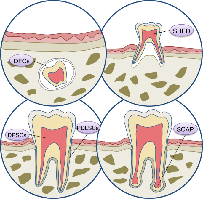


Stem Cell Based Bone And Dental Regeneration A View Of Microenvironmental Modulation International Journal Of Oral Science


Jaypeedigital Ebook Reader
Both materials will be applied in intrabony defects during periodontal flap surgery Followups will take place at 7 and 14 days post surgery, and at 3, 6, 9, and 12 months The primary aim is to compare the effectiveness of the test product with the control in the treatment of 1 and 2wall intrabony periodontal defects on the amount of boneMany times, more complex bone defects are encountered, where a threewall defect in the apical portion becomes two or one wall defect in the coronal region This is then referred to as a combined osseous defect One wall defect Two wall defect Three wall defect Clinical photograph of a one wall defect (Courtesy Dr Amit Wadhawan)The distribution of location and defect types for implants with 2, 3, or 4wall bone defects are presented (Table 1) Data on clinical assessments performed immediately before and during surgical procedures are presented for defects located in the maxilla or the mandible (Table 2)


Pathophysiology Of Periodontal Disease Revise Dental


Experimental Animal Models In Periodontology A Review
There are several published classification systems for acetabular bone defects in THA 1, 3, 5, 11, 12, 19, Although similarities exist between classification systems, each one has a unique grading scale ranging from mild to severe bone defects Precise planning of the operative procedure is indispensableBone loss in oblique direction Base of the defect is located apical to the surrounding bone Intra bony defectsall vertical defects three wall defect Vertical defects distal and mesial surface of molars Three wall three wall present intrabony defectnow all vertical defects are know was intrabony defects Mesial surface of second and third molar One wall defect hemiseptumClassification of Bone defects One wall defect – usually only one interdental wall remains and is called hemi septum if remaining wall is proximal Poor prognosis for periodontal regeneration since it is difficult to stabilize the graft material to be used in its proper place
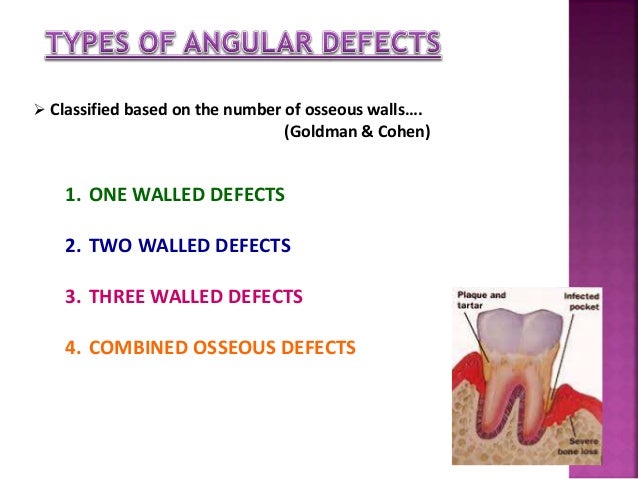


Patterns Of Bone Destruction In Periodontics


Classification Scheme For Intrabony Defects Figure 1 Reproduced Download Scientific Diagram
The bone destruction patterns that occur as a result of periodontal disease generally take on characteristic forms This Xray film displays a horizontal defect This Xray film displays two lonestanding mandibular teeth, #21 and #22 the lower left first premolar and canine, exhibiting severe bone loss of 3050%Molar teeth with three or more bony wall defects are most commonly lost if timely intervention is not done Etiology of intrabony defect involvement includes local environmental factors such as tooth position which contributes to food impaction, plaque accumulation, angular position, or position of the tooth with respect to the alveolarCase 2 A wide 3wall intrabony defect on the distal aspect of tooth #30 This defect was regenerated successfully using autogenous bone harvested from the adjacent edentulous ridge in combination


Periodontal Bone Defects Botiss Campus


Jaypeedigital Ebook Reader
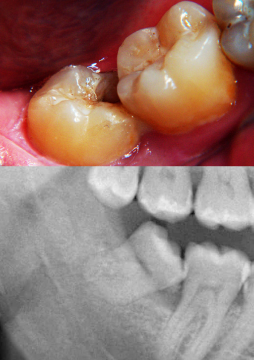
And none at the medial thirdOne wall nobly defect , Two wall bony defect, Three wall bony defect, Combined type /*นับจำนวนตาม bony wall ที่เหลืออยู่ */For #16, which exhibited a 2 to 3wall vertical bony defect and class III (mesiodistal) furcation involvement, bone graft was scheduled ncbinlmnihgov Tunnel preparation was performed on #46 as it had a 2wall vertical bony defect and Degree 3 furcation involvement


Classification Scheme For Intrabony Defects Figure 1 Reproduced Download Scientific Diagram


Patterns Of Bone Loss In Periodontal Diseases Periodontitis And Mechanism


Clinical And Radiographic Evaluation Of Platelet Rich Fibrin As An Adjunct To Bone Grafting Demineralized Freeze Dried Bone Allograft In Intrabony Defects Atchuta A Gooty Jr Guntakandla Vr Palakuru Sk Durvasula S Palaparthy R


Periodontal Regeneration Registered Dental Hygienist Rdh Magazine


Http Www Ijss Sn Com Uploads 2 0 1 5 Ijss Jun Oa59 17 Pdf


Periodontal Bone Defects


Bone Loss


Www Ecronicon Com Ecde Pdf Ecde 18 Pdf


p Onlinelibrary Wiley Com Doi Pdfdirect 10 1902 Jop 1967 38 6 Part1 455


Treatment Of Intrabony Defects With Modified Perforated Membranes In Aggressive Periodontitis Subtraction Radiography Outcomes Prognostic Variables And Patient Morbidity Springerlink


Recent Advances In Periodontal Regeneration A Biomaterial Perspective Sciencedirect


Q Tbn And9gctrozqhd8a Y4jijakzy08r4o0iwyhd8czl6q1pm8voulybvckp Usqp Cau


Extraction And Socket Grafting Part 2 Extraction Site Healing


Tha Revision Recon Orthobullets


Regenerative Techniques In Oral And Maxillofacial Bone Grafting Intechopen


Osseous Resective Surgeries Periobasics Com


Good Morning Ppt Download


27 Bone Defects And Furcation Lesions Pocket Dentistry


Interdisciplinary Management Of An Isolated Intrabony Defect


Intrabony Defects T2 6 Flashcards Quizlet


Acetabular Reconstruction Classification Of Bone Defects And Treatment Options Musculoskeletal Key


Extraction And Socket Grafting Part 3 Socket Grafting Protocol


Periodontal Regeneration Intrabony Defects Practical Applications From The p Regeneration Workshop Reynolds 15 Clinical Advances In Periodontics Wiley Online Library


Jaypeedigital Ebook Reader


Bone Defects In Periodontal Disease Foundations Of Periodontics Bone Defects Statistics


Periodontal Defects And Regenerative Success


Clinical And Radiographic Evaluations Of Periodontal Intrabony Defects Treated With Enamel Matrix Derivative A Report Of Two Cases Medcrave Online


Ppt Bone Defect Powerpoint Presentation Free Download Id


Mechanism And Patterns Of Bone Loss Ppt Download


Intrabony Defects T2 6 Flashcards Quizlet


The Image And Radiographs Of Three Types Of Defects W Wall Ddr Download Scientific Diagram


Bone Loss


Periodontal Osseous Defects A Review Semantic Scholar


Bone Destruction Patterns In Periodontal Disease Periodontal Disease


Interdisciplinary Management Of An Isolated Intrabony Defect


Patterns Of Bone Loss In Periodontal Diseases Periodontitis And Mechanism


Classification Scheme For Intrabony Defects Figure 1 Reproduced Download Scientific Diagram


Pdf Management Of A One Wall Intrabony Osseous Defect With Combination Of Platelet Rich Plasma And Demineralized Bone Matrix A Two Year Follow Up Case Report Semantic Scholar


Evaluation Of The Prevalence And Distribution Of Bone Defects Associated With Chronic Periodontitis Using Cone Beam Computed Tomography A Radiographic Study


Plos One Long Term Observation Of Regenerated Periodontium Induced By Fgf 2 In The Beagle Dog 2 Wall Periodontal Defect Model


Recent Advances In Periodontal Regeneration A Biomaterial Perspective Sciencedirect


Bone Loss


Improved Long Term Treatment Outcomes Of Teeth With Deep Pockets And Reduced Periodontal Support By Periodontal Regenerative Surgery Oral Health Group


Periodontal Regeneration Intrabony Defects Practical Applications From The p Regeneration Workshop Reynolds 15 Clinical Advances In Periodontics Wiley Online Library


Improved Long Term Treatment Outcomes Of Teeth With Deep Pockets And Reduced Periodontal Support By Periodontal Regenerative Surgery Oral Health Group


Economically And Clinically Leveraging Implantology In Your Solo Practice Dso Or Ddso Innovations In Immediate Implants Dental Economics


Bone Defects And Furcation Involvement Part 1 Intelligent Dental


The Use Of Augmented Antibiotic Loaded Cement Spacer In Periprosthetic Joint Infection Patients With Acetabular Bone Defect Journal Of Orthopaedic Surgery And Research Full Text


Improved Long Term Treatment Outcomes Of Teeth With Deep Pockets And Reduced Periodontal Support By Periodontal Regenerative Surgery Oral Health Group


The Periodontal Endodontic Relationship What Do We Know Intechopen


A Intraoral View Showing Defect In The Mesial Wall Of Open I


Q Tbn And9gcrlwkxb0xadxunfl2wlzoa0ubtvtrneqbzc0mxp8hpv7l Dj8 J Usqp Cau


Periodontal Disease Wikipedia


Jaypeedigital Ebook Reader


Patterns Of Bone Destruction In Periodontics


14 Periodontal Surgery Pocket Dentistry
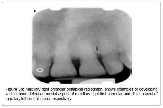


Different Radiographic Modalities Used For Detection Of Common Periodontal And Periapical Lesions Encountered In Routine Dental Practice Omics International
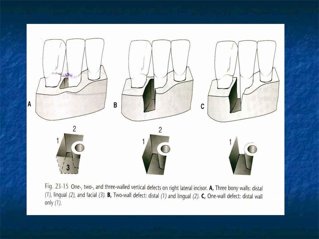


Bone Loss Patterns Of Bone Destruction Ppt Video Online Download


Jaypeedigital Ebook Reader


Management Of Intrabony Defects In Periodontal Disease Dental Update


Orthopedic Implants And Devices For Bone Fractures And Defects Past Present And Perspective Sciencedirect


Www Codsjod Com Doi Cods Pdf 10 5005 Jp Journals 0028


Osseous Resection With Apically Positioned Tooth Structure


Www Ecronicon Com Ecde Pdf Ecde 18 Pdf


Smile Dental Magazine Dentists Reference On The Web
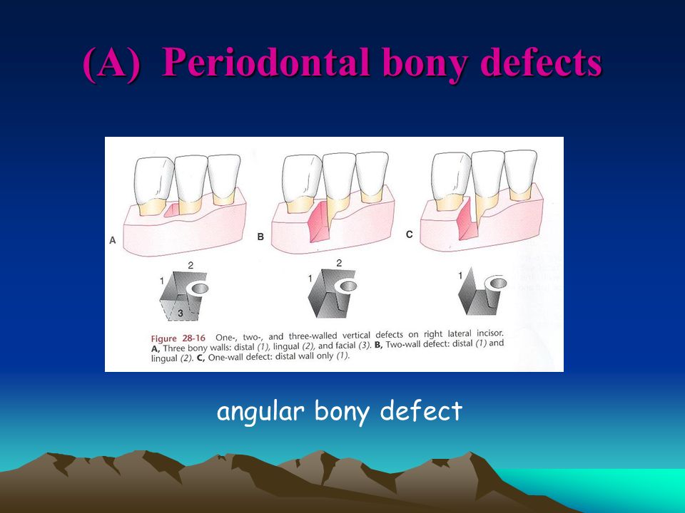


Ren Yeong Huang Dds Phd Ppt Video Online Download
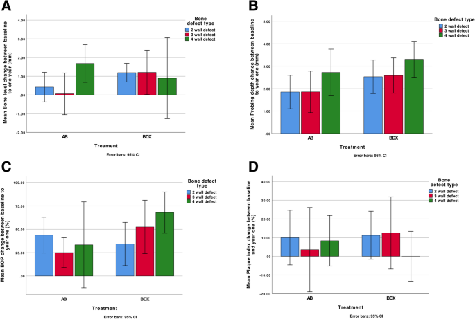


Impact Of Bone Defect Morphology On The Outcome Of Reconstructive Treatment Of Peri Implantitis International Journal Of Implant Dentistry Full Text


Classification Scheme For Intrabony Defects Figure 1 Reproduced Download Scientific Diagram


Www Dentaltown Com Images Dentaltown Magimages 1110 Dtnov10pg54 Pdf


Impact Of Bone Defect Morphology On The Outcome Of Reconstructive Treatment Of Peri Implantitis International Journal Of Implant Dentistry Full Text


Http Www Ijss Sn Com Uploads 2 0 1 5 Ijss Jun Oa59 17 Pdf


Titanium Mesh For Bone Augmentation In Oral Implantology Current Application And Progress International Journal Of Oral Science


Management Of Peri Implant Infections Vandana K L Dalvi P Nagpal D J Int Clin Dent Res Organ


Intrabony Defects T2 6 Flashcards Quizlet


コメント
コメントを投稿